

How the ECG is made: the process of performing a cardiogram
Author Ольга Кияница
2018-05-15
Method of ECG
The principle of the ECG is based on the ability of the device to capture the electrical impulses that arise in the heart and, after processing, record them in a graph.Further, the results obtained are interpreted by a specialist in functional diagnostics, and the attending physician can use the data to make a diagnosis.

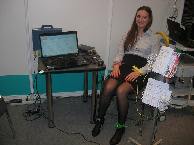
The electrocardiogram is performed with the help of a special apparatus - an electrocardiograph. Its main nodes are:
- galvanometer;
- lead switch;
- system for amplifying captured signals;
- recording device.
During the ECG removal procedure, electrodes placed at certain points capture the electrical impulses that arise in the heart and transmit them through the galvanometer to the recording device. Further, the recorders display the information received on a paper moving at a certain rate in the form of graphs that show the functioning of different parts of the heart.
ECG charts are recorded on millimeter paper in the form of lines of teeth of different sizes. These dimensions depend on the strength of the signal that is given by one or another part of the heart.
Video: Recording an electrocardiogram
The results of the study are described by a specialist who compares these norms with the results obtained. In the interpretation, the doctor of functional diagnostics indicates the revealed deviations, according to which the attending physician of the patient makes a diagnosis.
When is electrocardiography necessary?
Indications for the appointment of an electrocardiogram are the following complaints of the patient or symptoms:
- dyspnea;
- uncomfortable and painful sensations in the chest;
- frequent stress;
- tachycardia or bradycardia;
- signs of arrhythmias;
- overweight;
- suspicions of hypertension and other manifestations of cardiovascular diseases;
- preparation for a surgical operation or for some types of diagnostic studies.
For prophylactic purposes, the ECG should be administered to all individuals older than 40 years once a year. At a more mature age, the study should be performed once a quarter. For people suffering from cardiac and vascular pathologies, an electrocardiogram is carried out as often as necessary, that is, at the time that the doctor, guided by the data on the existing disease, has determined for them.
How is the cardiogram read and what determines the ECG?
Conclusion on the ECG is performed by a physician of functional diagnostics and a cardiologist. The description of the study includes several stages:
- an estimation of age and sex of the patient - from these data interpretation of results by age norms depends;
- determination of the amplitude, duration of the teeth and intervals of the cardiogram - analysis of the P wave and the duration of the P-Q interval, the QRS ventricular complex, the ST segment, the T wave, the Q-T interval;
- characteristic of the rhythm - in arrhythmias, an irregular rhythm is detected, an increase in the frequency of heart contractions of more than 100 beats per minute, a violation of the conductivity of an electric pulse through a conducting system, or a nonsinus rhythm;
- the presence of focal changes in the heart muscle - when identifying zones of myocardial infarction, the localization of the focus of necrosis and its prevalence is determined;
- manifestations of hypertrophy of these or those parts of the heart - an increase in the size of the heart muscle is manifested on the ECG increased electroactivity of the enlarged chamber, ischemic or dystrophic processes in the myocardium, or by the slow spreading of the electrical impulse in the wall of the hypertrophied cardiac chamber.

In addition to the above changes in the heart, an electrocardiogram can help evaluate the features of the organ position, signs of myocarditis, pericarditis, the condition of the implanted pacemaker, electrolyte exchange disturbances, and other abnormalities.
After the analysis of all the data obtained, a conclusion is formed. If possible, a comparison of the ECG with the previously recorded cardiogram charts is performed, that is, the results are monitored in dynamics.
Normal ECG parameters are as follows:
- rhythm - regular and regular, with a source in the sinus node;
- heart rate - determined by measuring the distance between a number of adjacent teeth R (interval R - R), assess the indicator according to age (for adults and healthy people up to 50 years - 60 - 90 beats per minute);
- the electrical axis of the heart - normally the alpha angle is 40 - 70 °.
Video: ECG norm. All intervals and prongs: p, QRS, T, PR, ST
Features of cardiography children
ECG can be administered to children of any age. When evaluating the results, the doctor is necessarily guided by age characteristics and, if necessary, complements the study with other techniques (eg, Echo-KG, cardiac CT, etc.).
ECG in children has its own specificity in each age period, since the heart is affected by many factors depending on age and growth: changes in the number of cardiac contractions, the influence of the vegetative and endocrine systems, anatomical positions of the heart in the chest, changes in the speed of pulse propagation and etc.
The following clinical cases may become a reason for the appointment of an ECG to a child:
- noises in the heart;
- heart rhythm disturbances;
- presence of even minor dyspnea;
- pathology of other organs;
- presence in the history of severe infectious diseases;
- detection in a patient's history of relatives with severe cardiac pathologies;
- the need for surgical treatment;
- preventive examination in sports.
Performing ECG for children who have cardiac and vascular diseases or are at risk of their occurrence should be conducted at least once a year.
Of particular importance are the features of the preparation of the child for research. During the recording of the cardiogram chart, a small patient should be calm and still, and this effect can not be achieved with all children. For older children, parents and a doctor explain the need for adherence to established rules, and younger children are encouraged to distract themselves with toys or the so-called "play in the hospital." Such psychological tricks almost always help to achieve the patient's necessary calmness for investigation, and the results obtained will be reliable. If the child can not behave calmly during the procedure, then parents are advised to come another day to record the ECG.
Features of the passage of ECG in women during pregnancy
Conducting ECG is absolutely safe procedure and not dangerous for pregnant women. That is why women should not refuse to perform such a study, allowing time to identify various abnormalities in the work of the heart, experiencing an increased load during the gestation of the fetus. During pregnancy, the ECG procedure is prescribed 2 times, besides this, the electrocardiogram is performed not only by the woman, but also by the fetus - such a study is called CTG (cardiotocography).
When interpreting the results, the doctor necessarily takes into account the peculiarities that occur in the body of a woman during pregnancy (especially in her late periods) and affect the nature of the cardiogram schedule:
- displacement of the electric axis to the left;
- increase in the number of heartbeats;
- negative T wave in the third and fourth lead;
- shortened interval PR;
- pathological prong in Q in the third lead and aVF (right arm).
How to do an electrocardiogram?
Preparation before ECG, to obtain reliable diagnostic data, is carried out as follows:
- The last meal should be 2 hours before the test.
- Before the procedure, the patient must abandon physical exertion, smoking, drinking cold water, alcohol and caffeine-containing products and beverages.
- On the day of the study, the patient should avoid applying creams and other fat-based cosmetic products to the body, since such substances can interfere with sufficient contact between the electrode and the skin.
- Before visiting the ECG cabinet, the patient should wear comfortable, breath-free clothing that easily stretches, rises from the limbs or is removed. From the neck and hands should be removed metal jewelry (chains, bracelets, necklaces, etc.).
- Take the previous ECG or Echo-CG results with you. Using this data, the doctor will be able to analyze the dynamics of the disease and this will improve the reliability of the final diagnosis.
- The patient should be calm before removing the ECG. To achieve this emotional and physical balance, he should lie down or sit on the couch 10 - 15 minutes.
- For a patient who has shortness of breath, the examination should not be performed in the prone position (as usual), but in the sitting position.
- The room in which the electrocardiogram is performed should be ventilated and with an optimal temperature regime, since a cold sensation during the procedure may negatively affect the reliability of the results of the study.
An electrocardiogram is performed in the following sequence:
- The patient unbuttons or removes clothing for convenient attachment of electrodes in the points required for the study and lies on the couch.
- Electrode attachment areas are treated with alcohol to degrease the skin, and then a special gel is applied on them.
- The doctor turns on the device and for a certain time receives the schedule necessary for the subsequent decoding.Usually the study takes no more than 15 - 20 minutes.
After the ECG, the patient can wait for the results of the survey to be described or sent to his or her healthcare provider.If serious violations have been detected in the electrocardiogram schedule, then the patient's doctor should immediately be connected to the patient's examination, who can correctly assess all the data and prescribe the treatment appropriate for the detected clinical situation.
In most cases, the ECG is well tolerated by patients. Sometimes on the place of fixing the electrodes during the study, there are sensations of weak and hardly perceptible electrical discharges, and in some patients after the examination a small rash appears on the skin, which is eliminated independently in a short time and does not require treatment.
The ECG carries no danger to the health of the patient, since during the examination, X-ray irradiation and other factors harmful to the organism are not applied. If necessary, this method of examination can be repeated, even at short intervals.
Conclusion
The ECG makes it possible to detect abnormalities in the electrical activity of the heart, arising from arrhythmias, organic myocardial lesions, metabolic disorders of potassium, calcium and magnesium, pain in the heart and increasing the thickness of its chambers. This informative study is conducted in almost every clinic, is completely safe and can be prescribed to people of any age category and pregnant.
Similar articles

The heart belongs to vital organs, therefore without it it would not have been possible for a single person to exist. The structure, as well as the appearance of the heart, is rather diverse, but quite capable of logical explanations. A proper understanding of how the human heart looks like a healthy estimate of the body’s overall capacity.
Modern technologies allow the most thorough study of various organs and tissues, including the cardiovascular system. Without timely and correct diagnosis sometimes it is very difficult to put the exact diagnosis. At the same time, it is extremely important to do everything in time to ensure effective treatment after the study.

There are a number of serious pathologies of the circulatory system that can lead to serious consequences. One of them is a vascular thrombosis, in which arteries, veins, and capillaries can be affected. But the most dangerous condition is the formation of blood clots inside the heart. Such a violation must be eliminated as soon as possible, otherwise serious consequences may arise.
Вышеперечисленные и многие другие изменения, выявленные при расшифровке ЭКГ (электрокардиограмма), являются признаками сердечной патологии. Поэтому сделать кардиограмму сердца необходимо для раннего выявления сердечных заболеваний, пока ещё возможна эффективное лечение и профилактика осложнений. Электрокардиограмма позволяет поставить или подтвердить диагноз ишемической болезни сердца, гипертонической болезни, инфаркта миокарда, аритмии и др. Сделать кардиограмму сердца необходимо и в рамках профилактического обследования, а также женщине при подготовке к беременности. При сердечном приступе обязательно сделать кардиограмму, и чем быстрее, тем лучше.