

Intracardiac thrombi and what they threaten
Author Ольга Кияница
2017-10-30
Thrombosis is a pathological condition related to diseases of the cardiovascular system. During the formation of thrombus, bleeding through the vessels (arteries, veins and capillaries) worsens, resulting in ischemic areas, up to necrosis. Severe consequences are determined in those cases when a blood vessel clogs a thrombus, but even the cessation of blood flow in a small capillary can lead to unpleasant complications.
In the cavities of the heart, especially in the atria, thrombi can also form. When they are detached from the endocardium and begin to move with the flow of blood, they are called emboli. Thrombotic clots can get into various organs and even the brain, but most often thrombosis of the pulmonary artery occurs, which leads to acute heart failure.
After determining the primary location of the thrombus, it is important to establish the path of the embolus. This will determine the most likely target organs. If the embolus enters the circulatory system from the lower hollow veins and the right atrium, then it clogs the pulmonary artery. Intra-cardiac thrombi can be directed both to the vessels of the brain and to the blood supply system of the lungs.
Video What is a thrombosis, and why is it dangerous for the heart?
The causes of the formation of intracardiac thrombi
The formation of thrombi causes considerable damage to the circulatory system. Awareness of the causative factors leading to thrombosis helps to choose the right treatment for pathology.
The main triggers of intracardiac thrombus development are:
- insufficient speed of blood flow;
- violation of the integrity of the endocardium and valves, which leads to the formation of vortexes of blood flow;
- increased blood coagulability.
These pathological processes can occur with various diseases. Most often, cardiac defects contribute to this, especially mitral stenosis, which is often formed with rheumatism. As a result, the thrombus forms in the left atrium.
Saciform protrusions, called aneurysms, appear against the background of thinning of the heart wall. Especially this is promoted by the transferred transmural myocardial infarction, which through time is replaced by scar tissue. Most often, the right and left ventricles are involved in the pathological process.
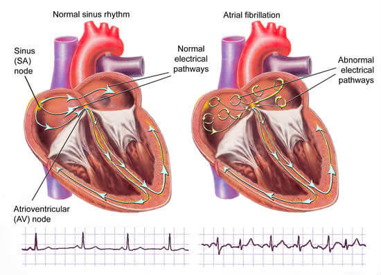
In some cases, the heart muscle begins to contract unevenly. This contributes to the occurrence of arrhythmias, which in turn provoke the development of intracardiac thrombi.
Increased synthesis of fibrin, which plays an important role in the formation of blood clots, is observed in a number of diseases. In particular, ESR increases with angina, ARVI, inflammatory lung diseases, kidneys and other organs. If there is no timely help in such diseases, then the risk of thrombus formation and thromboembolism of various vessels is significantly increased.
Features of thrombi
There are several forms of blood clots, which differ from each other in appearance and structure. Depending on the type of vessel, the thrombus can be:
- Red (formed in the veins and consist of erythrocytes, fibrin, leukocytes and platelets);
- white (arise in the arteries and consist of the same cells, except for erythrocytes);
- hyaline (found in capillaries of different caliber and contain protein components instead of fibrin).
Intracardiac thrombi is characterized by a mixed structure. On appearance they are mottled, so the second common definition is layered blood clots. The main parts of their structure are the head, body and tail. The initial element is similar to a white blood clot, and the last one is red. It is the "white" part in most cases that the thrombus is attached to the endocardium (inner shell of the heart).
Intracardiac thrombi can be mobile and immobile. The former are more dangerous, since they can move through the ventricles and atria, often leaving the general circulatory system. The main place of their formation is the left atrium.
Depending on the ratio of the thrombus to the heart cavity, the occluding and parietal thrombi are isolated. The first often cause the death of the patient, because they block the path of blood flow due to the complete overlap of the lumen of the cavity. The second type of thrombus is more often detected and often occurs against the background of inflammatory processes that pass from the endocardium to the valve structures. Due to their attachment to the auricles for blood flow there is free space, but it can be significantly narrowed, which also affects the course of the disease. Parietal thrombi are mainly formed in ischemic heart disease, chronic heart failure, aortic aneurysm.
Symptoms of intracardiac thrombosis
The clinical picture depends on the form and location of localization of thrombus formation:
- With a fixed thrombus, in rare cases, accelerated heart rate and dyspnoea in a sitting position are determined.
- The movable thrombus in the left atrium provokes the appearance of such symptoms as the appearance of cold sweat, debilitating attacks of palpitation, loss of consciousness, marked pallor of the skin, against which the cyanosis of the fingers and lips is noticeable.
- For a thrombus spherical shape or when it is attached to a pedicle, the appearance of a poor state of health in sitting position is typical. The appearance of this sign is explained by the lowering of the thrombus and the occlusion of the venous aperture. When such a patient takes a horizontal position, it becomes much easier.
In a number of cases, there is a reflex spasm of cerebral vessels due to increased excitability of the region of the atrial auricles. A similar condition can provoke a fainting or fainting condition.
Acute disorders of the various organs can indicate the separation of the thrombus in the heart cavity and its entry into the general system of blood flow:
- acute ischemic stroke, often manifested by changes in vision and speech, as well as paralysis, occurs due to thromboembolism of cerebral vessels;
- acute myocardial infarction is expressed by severe pain in the heart, shock state and often develops against the background of blockage of blood flow in the coronary artery;
- blockage of the jugular vein on the neck is often manifested by a sharp dizziness, very severe headache;
- embolism of the arteries of the kidneys is accompanied by severe pain that is localized in the lumbar region and can be accompanied by a urinary tract disorder;
- the cessation of blood circulation in the mesenteric vessels leads to severe peritonitis complicated by necrosis of intestinal loops, and is expressed by palpable pains, swelling of the intestine;
- occlusion of the vessels of the lower and upper extremities leads to the appearance of severe pallor, cooling of the limbs, increased risk of gangrene with subsequent amputation.
Diagnosis of intracardiac thrombi
In the case of intracardiac thrombi, electrocardiography is not very informative. This is due to the ability of the technique to evaluate more the work of the conduction system of the heart than its internal structure.
The method of selection in the definition of intracardiac thrombi is ultrasound examination of the heart.
Less informative is considered dopplerography, so if possible and the presence of a characteristic clinic, patients with suspicion of blood clots inside the heart cavities immediately go to ultrasound. During the examination, the structure of the atria and ventricles is visualized, in some cases it is possible to determine thrombi of relatively small size.
Video Thrombuses up to 4.0cm x 2.5cm. in the left atrium (LP transverse size 10.0cm)
Treatment of intracardiac thrombi
Patients with thrombosis inside the heart should be operated in a timely manner, because only this method allows to remove from the cardiac cavity thrombus formation. If one uses only conservative therapy, then with her help it is possible to prevent the appearance of thrombi.
Most often, intracardiac thrombosis introduces the patient into a critical condition, when the operation can save his life. After its successful implementation, as a rule, the prevention of recurrence of thrombus should be performed. For this, the correction of clotting factors is carried out and the underlying disease that provokes thrombus formation is necessarily treated.
In complex therapy, patients need rheumatism, chronic heart failure, high blood pressure, blood diseases, which are accompanied by an increase in coagulation rates.
Normal blood viscosity is maintained by taking drugs from the following pharmacological groups:
- anticoagulants (heparin, hirudin, warfarin, dicumarin), which are of indirect and direct action., help to correct factors of blood coagulability;
- antiaggregants (acetylsalicylic acid, dipyridamole, iloprost) - affect the ability of platelets to stick together, which in turn prevents thrombosis.
With all the detailed questions, you should contact a cardiologist who, after examining the patient, will suggest a tactic for treating intracardiac blood clots.
Similar articles

Thrombosis is a process of blood clotting, the result of which is a blood clot in the blood vessel. This clot may block or impede blood circulation in the affected area, as well as cause serious complications. Especially dangerous is a thrombus that moves to the critical part of the circulatory system, such as the brain or lungs.
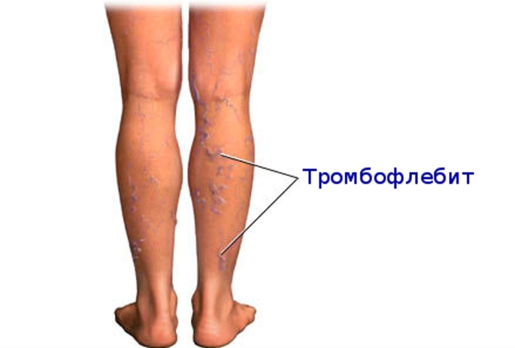
Relatively favorable thrombophlebitis may overnight become dangerous to the patient's life. Much depends on the localization of thrombophlebitis, but in any case, timely treatment should be performed. It is conducted by a phlebologist or, at worst, by a surgeon.

The main driver of the rhythm of the heart, sinus node, has an interesting history of discovery and a number of amazing features in the structure and functioning. From the coordination of the work of this part of the heart depends the overall activity of the whole organ, therefore, with dysfunction of the sinus node, treatment is always carried out, otherwise there is a risk of fatal outcome.