

Retinal angiopathy
Author Ольга Кияница
2018-06-26
Angiopathy of the retina (AC) - a pathological change in the capillaries and arteries of the eye. They can be thickened or, alternatively, enlarged, wriggled, tapered, which leads to a violation of the blood supply to the eyes. AC is not considered an independent disease, because it mainly occurs against the background of other pathological conditions associated with the heart, the brain, etc.
Angiopathy (Novolat, angiopathia, dr.-Greek ἀγγεῖον - vessel and πάθος - suffering, disease, synovial vasopathy) is a general term for the disease of blood vessels (arteries, veins and capillaries).
The development of angiopathy is most often associated with a disorder of nervous regulation, which contributes to the development of dystonia, unstable spasms and paresis of the vascular network. First, the changes are reversible, but with a prolonged course of the disease and the absence of treatment can become irreversible.
Video: Angiopathy of the retina is what it is in adults and treatment
Causes of retinal angiopathy
Ophthalmologists in most cases consider retinal angiopathy a symptomatic manifestation, that is, a sign of other disorders, among which the following are most often highlighted:
- Hypertension, in which arteries are exposed to the negative influence of high blood pressure
- Low blood pressure
- Diabetes mellitus, which is characterized by the defeat of various organs and systems of the body, including the eyes
- Atherosclerotic vascular lesions
- Head trauma, which greatly increases intracranial pressure, or the spine (especially the cervical spine), which often leads to a violation of blood flow
- Osteochondrosis of the cervical spine
- Hereditary vascular diseases
- Harmful habits (binge, smoking, drugs)
- Work on harmful to health production
- Diseases of the blood
- Age over 30 years
Types of retinal angiopathy
In modern clinical medicine, six forms of the disease are distinguished, which basically differ from each other in the cause of the onset.
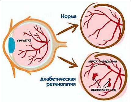
1. Diabetic
Many diabetics have this form of angiopathy. It often develops in 5-10 years of the course of the underlying disease.Patients may be affected by larger vessels, in this case they speak of macroangiopathy, in addition to the affected capillaries, when doctors note the presence of microangiopathy.
2. Hypertensive
With hypertension, large vessels suffer from high blood pressure, while the retinal arteries begin to contract against the background of the compensatory mechanism. Such changes disturb the blood flow in the organs of vision.
3. Hypotonic
With prolonged course of hypotension, the retinal vessels widen compensatoryly, which leads to visual impairment, dizziness and headache.
4. Congenital
It is determined immediately after the birth of the child. It is more common in premature babies who suffer from underdevelopment of blood vessels.
5. Traumatic
It is characterized by rupture of the vessels of the retina caused by increased physical work or damage to the tissues of the head, brain, face, other parts of the body. Sometimes, during the birth, retinal rupture can also occur, which results in complete or partial blindness.
6. Youth
The pathology manifests itself as a hemorrhage into the retina, caused by the fragility and fragility of the capillaries.There is a rare disease and until the reasons for its development are not fully understood.
Angiopathy during pregnancy
Separately it is necessary to consider angiopathy during pregnancy. As a rule, it is not dangerous for a woman. After childbirth can self-resolve after about 2 - 3 weeks. The emergence of pathology is often associated with increased blood circulation and excessive proliferation of blood vessels.
If retinal angiopathy was diagnosed before the onset of pregnancy, then the treating ophthalmologist will certainly notice the violations and take measures to prevent possible problems and unpleasant consequences.
Sometimes angiopathy in pregnancy is complicated by pathological conditions, which endanger the life of a woman and a baby. In such cases, delivery by a caesarean section may be required.
Signs of angiopathy of the retina
At the first stages of angiopathy is almost not manifested, which is dangerous for the overall health. Initial changes can be observed only by an ophthalmologist during the next examination of the patient. Some patients have the following symptoms:
- Unclear, reduced vision
- Sensation of pulsation in the eyeball
- Fatigue with hard eye work
- Eye swelling, inflammation of the eyeball and eyelid
- Discomfort and pain when touching the eyes.
With a sharp deterioration of vision, you do not need to delay a visit to the doctor, since the risk of developing blindness is high.
Diagnosis of retinal angiopathy
Angiography
Fluorescein and indocyanine green angiography are diagnostic tests that use special cameras to photograph structures in the back of the eye. These tests are very useful for detecting spasm or damage to the blood vessels that feed the retina. In both studies, the contrast agent is injected into the vein of the hand, which passes through the circulatory system and reaches the vessels of the retina, and then the deeper layers of tissue called the choroid. These tests are relatively harmless, because they are not associated with the use of X-rays or other harmful forms of radiation.
Fluorescein is a yellow dye that glows in visible light. Indocyanine is a green dye that fluoresces with invisible infrared light; so to determine it requires a special digital camera, sensitive to these rays of light.

Risks in angiography of the eye
Most often, the study does not cause side effects, but there is a possibility that the patient may have a reaction to contrast agents. Although fluorescein does not contain iodine and is safe for patients who suffer from allergies, indocyanine green is being done with iodine, so it should not be used in this category of patients.
Some people may experience mild nausea after injection of the dye, which usually passes quickly. In patients who are allergic to the dye, itching and skin rashes may occur. Very rarely there is a sudden and life-threatening allergic reaction, called anaphylaxis. This condition requires urgent medical treatment.
Additionally, it is possible to penetrate the dye into the skin at the injection site; this can cause some discomfort or discoloration of the skin for several days. Fluorescein dye also promotes the acquisition of urine orange color, can slightly discolor the skin for a short period of time.
Optical coherence tomography
Optical coherence tomography (OCT) is a diagnostic test that allows you to visualize and measure the thickness of the retina. OCT is very useful in detecting the swelling of the retina or the accumulation of fluid secondary to various states of the retina. The method provides very valuable information and is also useful for investigating the response to treatment.
OCT testing has become the standard for the evaluation and treatment of most retinal diseases. The survey takes only a few minutes. This test does not use radiation or X-rays.
Ultrasonography
Ultrasound (ultrasound) is a test that uses sound waves to assess the state of the eyes and the retina. If the doctor can not see the retina because of some opacity that blocks the visual image, ultrasound can be used to determine the general state of the retina. This method is commonly used to evaluate the retina in patients with dense cataracts or vitreous hemorrhage. Ultrasound is easy to use, while painless and does not require the use of any radiation.
Amsler's grid
This test is designed to detect anomalies in the macula, the area of reading the eye. This helps to diagnose many diseases at an early stage, when they can still be treatable. Ideally, the Amsler grid should be viewed daily. It is easier to remember such a thing if the grid is located where the person will necessarily see it, for example, on the refrigerator or mirror in the bathroom.
The grid consists of a pattern of vertical and horizontal lines with a dot in the center. You need to check each eye separately, closing the other eye. Hold the net about 45 cm from your face. It is necessary to look only at the central point, and, doing this, pay attention to whether all the lines are present.
You should also look for any areas where the lines are wavy or distorted. About those areas where the grid is absent or wavy, distorted lines that differ significantly from the baseline, should be reported immediately to the ophthalmologist.
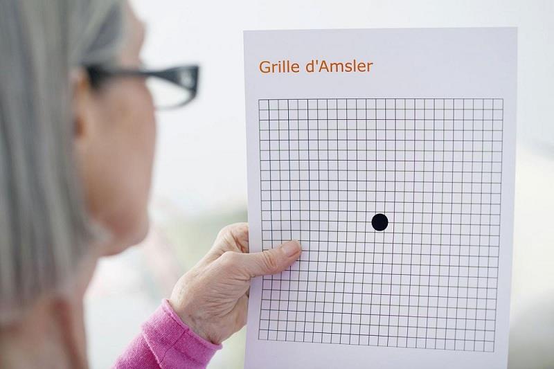
This relatively simple, inexpensive way of analyzing vision is used to detect changes in the central vision. Despite the fact that new technologies are being developed for earlier detection of problems with macular problems and are becoming more accessible, the Amsler grid remains a useful universal diagnostic tool.
Treatment of retinal angiopathy
Treatment of this disease is quite long and difficult. First the doctor finds out what causes angiopathy, and takes measures to eliminate them. For example, in determining a hypertension or hypotension in a patient, the therapy is aimed at normalizing blood pressure, and in diabetes, proper nutrition and sometimes insulin therapy is extremely important.
In patients with a disturbed cardiovascular system and a related complication in the type of angiopathy of the retina, special drugs are used that activate blood circulation in the eyeball, improve the trophism of tissues and saturate them with sufficient oxygen. In such cases trental and mildronate are very effective. It is also important to adhere to the principles of proper diet and avoid foods rich in cholesterol. Positive, but moderate exercise exercises have a positive effect on health. Also, special vitamin complexes will be useful.
In the presence of serious lesions or occlusion of small ophthalmic vessels, laser therapy methods are used.
Video: Angiopathy of the retina is what it is in adults and treatment
Prophylaxis of retinal angiopathy
To prevent the development of angiopathy, first of all, the treatment of major diseases is carried out. Also adhere to the following recommendations:
- With diabetes : you need to keep glucose levels under control, adhere to dietary intake, take antidiabetic medications, and give up harmful habits.
- With hypertension and hypertension : take timely prescribed medicines, do not overwork physically, do not succumb to excessive cold or heat, do not smoke, do not drink alcohol and do not abuse coffee.