Aortic valve replacement: cardiac surgery
Author Ольга Кияница
2018-09-24
Aortic valve replacement is a method of surgery that is used to treat a cardiac disorder caused by damage to the aortic valve. In most cases, in the absence of surgical exposure to heart disease, severe disorders occur, leading to changes in the functioning of internal organs.
The aortic valve helps regulate blood flow from the heart to all organs and tissues. The valve also allows blood to flow in one correct direction.
When the left ventricle contracts, the aortic valve opens, allowing blood to enter the aorta through the aortic opening. During ventricular relaxation, the aortic valve closes, preventing blood from returning from the aorta. As a result, oxygenated blood enters the brain and all internal organs belonging to the large circulation.
Video: Replacing the aortic valve
History
Early surgical approaches to aortic valve disease were limited by the need to work with a constantly contracting heart. In the 1950s, the Hufnagel valve was implanted in the descending part of the thoracic aorta in patients with aortic insufficiency.
The first successful replacement of the aortic valve was registered in 1960 under the leadership of Harken.
Introduction to the practice of the presented method proceeded slowly, based on the limitations of the available interchangeable valves and the relatively primitive methods of protecting the heart during surgery that were available at that time. With the gradual introduction into practice of mechanical heart valves, the development of cardiopulmonary bypass surgery (cardiopulmonary bypass) and cardioplegia, which make it possible to safely stop the heart during surgery, aortic valve replacement has become available for patients with severe aortic insufficiency or regurgitation.
Description of aortic valve defects
Diseases (defects) of the aortic valve occur when, for various reasons, the structure of its cusps or aortic opening is disrupted. In particular, the following aortic valve defects are distinguished:
- Aortic valve stenosis (narrowing of the aortic valve that restricts blood flow).
- Smoothing of the aortic valve (valve insufficiency, which leads to incomplete closure of its cusps).
- Regurgitation of the aortic valve (the blood that is released into the aorta immediately returns to the left ventricular cavity, due to which the heart stops normal contraction).

Facts about aortic valve disease:
- They kill 25,000 people a year.
- It is determined in 7% of the world's population over the age of 65.
- 2 years after the diagnosis of severe aortic stenosis in patients who did not receive treatment, the survival rate reaches 50%.
It is when the aortic valve does not work properly, the hemodynamics becomes difficult and the blood does not flow well to the organs and tissues. With defects, the load on the heart also increases, which has a further negative effect on the maintenance of uniform circulation of blood throughout the body.
Symptoms of aortic valve defects
In some patients, significant changes are not observed for many years, while in other patients the symptoms manifest themselves very quickly and markedly. The most common symptoms of aortic valve disorder include:
- Chest or chest pain.
- Arrhythmia (sinus tachycardia, atrial flutter).
- Dyspnea (respiratory failure).
- Disorder of consciousness.
- Fainting
- Fatigue.
- Heart failure.
- Heart failure.
Diagnosis of aortic valve disease
With an objective examination of the patient, sometimes the doctor can immediately establish the diagnosis of aortic insufficiency. In particular, there may appear cyanosis of the skin, earlobes and nasolabial triangle. When auscultation can also be heard changes by the type of weakening of the first tone at the apex of the heart, dancing carotid, etc.
Additional research methods:
- Electrocardiography (ECG).
- Echocardiography (echocardiography or ultrasound of the heart).
- X-ray.
- Doppler research.
- Phonocardiography.
Depending on the condition of the patient and the prognostic conclusion, aortic valve replacement may be recommended. This surgical procedure is very effective in treating severe valve lesions. A successful operation relieves or allows you to completely eliminate the painful symptoms due to the regulation of hemodynamics in the body. As a result, surgical therapy helps prolong the life and strengthen the dynamics of the heart muscle.
Aortic Valve Replacement Procedure
There are various factors that are taken into account when deciding on the need for a replacement procedure. Some of these factors are:
- Age of the patient.
- Health status.
- Concomitant diseases that may contribute to the progression of aortic valve disease.
Usually, doctors choose heart valve repair, because this method has the lowest level of risk. With the help of various ways of repairing the valves of the valves, the condition and functioning of the heart is improved. In addition, the need for anticoagulants (blood thinners), which are often recommended for many years after some types of valve replacement, is significantly reduced.
Difference between reconstruction and replacement of the aortic valve
In some patients, there is a violation of the structure of the aortic orifice, which leads to improper closing of the aortic valve cusps. In such cases, doctors suggest a reconstruction procedure. This decision mainly depends on the severity of the patient's condition, because valve defects can not always be “repaired”.
It is important to know that valve reconstruction is a more complex process compared to the replacement procedure.Only after careful research and meetings of doctors of various specializations can a decision be made to restore the damaged valve.
Aortic valve replacement can be done in two ways:
- Using open heart surgery.
- Through a transcatheter surgery (TAVR), that is, aortic valve replacement using a catheter.
During an open-heart surgery, a large incision is made in the patient's chest. During the execution of TAVR, all the required manipulations are performed through a small hole in the sternum region. As a result, the second option for replacing the aortic valve is less complex, more efficient and involves less risk than open-heart surgery. Only a doctor can decide which of the proposed options is the best, most effective and safe for a particular patient. In this case, much depends on the state of human health and his medical history.
Types of valves
There are two main types of artificial heart valve:
- Mechanical valves.
- Fabric valves.

Mechanical valves
Mechanical valves are designed to ensure that the patient can use them for a long time. Although mechanical valves are durable and usually represent a one-step solution, there is an increased risk of blood clots when used. For this reason, all patients who have been implanted with mechanical valves must take anticoagulants (blood thinners), such as warfarin, throughout their life. The result is a pronounced tendency to bleed. Also, the sound of the mechanical valves can be heard with the naked ear, which sometimes reduces the quality of life of the patient.
Fabric valves
Tissue heart valves are usually made from animal tissue, part of an animal heart valve, or animal pericardium tissue.The selected material is subjected to special treatment to prevent rejection and calcification.
There are alternatives to fabric material valves. In some cases, a homograft can be implanted - the human aortic valve.Homographic valves are provided to patients and are amenable to recovery after another person (a valve donor) dies.The durability of the valves by the type of homographs is comparable to valves of pork and bovine tissue.
Another procedure for aortic valve replacement is the Ross procedure (or pulmonary autotransplantation). In the Ross procedure, the aortic valve is removed and replaced with the patient’s own pulmonary valve. A pulmonary homograft (pulmonary valve taken from a deceased person) is then used to replace the patient’s own pulmonary valve. This procedure was first applied in 1967 and is used mainly in children, since the operation allows the patient's pulmonary valve (installed at the aortic site) to grow with the child.
Valve selection
Tissue valves tend to wear faster due to increased exposure to blood flow. This condition is especially true for active (usually younger) people. Today, tissue valves, as a rule, can last about 20 years, but they definitely wear out faster in young patients.
When the fabric valve fails and needs to be replaced, the person must undergo a second operation to install a new valve. For this reason, younger patients are more likely to have mechanical valves installed, which helps prevent the increased risk and associated inconvenience associated with the re-implantation of another valve.
Open heart aortic valve replacement
Replacement of the aortic valve is most often done through a medium sternotomy, that is, the implantation of the device is carried out by cutting the sternum. After opening the pericardium, the patient connects to the heart-lung machine. This device assumes the functions of breathing and pumping the blood necessary for the patient, while the surgeon replaces the heart valve.

After the patient is connected to all necessary equipment, the surgeon removes the patient's aortic valve through special incisions and a mechanical or tissue valve is placed in its place. As soon as the device is in place and the aorta is closed, the patient is disconnected from the heart-lung machine.
Transesophageal echocardiography (TE echoCG, ultrasound of the heart through the esophagus) can be used to check the correct functioning of the valve. Sometimes, after the operation, there are any complications, to prevent their development, sensors are installed in the necessary places, which allow, if necessary, to correct the work of the heart.Drainage tubes are also inserted to remove accumulated fluid from the chest and pericardium after surgery. They are usually removed within 36 hours, while sensors for stimulation are usually left until the person is discharged from the hospital.
Hospital stay and recovery
After replacing the aortic valve, the patient is most often in an intensive care unit for 12–36 hours. It is allowed to go home after about four days, without complications of the type of heart block.
Recovery from aortic valve replacement usually takes about three months if the patient is feeling well. At the same time, undergoing surgery it is not recommended to do hard work for 4-6 months, which allows avoiding damage to the sternum and divergence of the stitches.
Transcatheter aortic valve replacement
This minimally invasive surgical procedure allows the implantation of a new valve without removing the damaged one.Upon successful operation, the new valve after its installation begins to perform the functions of the old aortic valve. The operation can be called transcatheter aortic valve replacement (TAVR) or transcatheter aortic valve implantation (TAVI).

The principle of operation of the valve-in-valve
During the operation, a device, somewhat like a stent, is placed in the artery and then a replaceable device is delivered through the catheter to the location of the damaged valve, while in a completely disassembled form.
As soon as the new valve expands, it pushes the flaps of the old valve to the side, while the replacement device begins to perform work on regulating blood flow.
The procedure presented is innovative and approved by the FDA for people with symptomatic aortic stenosis, who are classified as intermediate or high risk patients for standard valve replacement surgery. Differences between conventional and minimally invasive methods of exposure are significant.
TAVR procedure
Regular valve replacement is done through open access to the heart, for which a sternotomy is done. As a result, the thorax is surgically opened for surgery. In contrast, the conventional TAVR or TAVI treatment method can be performed through very small holes that leave all the bones of the chest intact.
There are also certain risks when performing TAVR, but this method is a more affordable treatment option for people who previously could not undergo surgery. At the same time, an additional bonus is provided in the form of faster recovery in most cases. The experience of treating patients through the TAVR procedure can be compared to cardiac ballooning or angioplasty in terms of time for completion and recovery. As a result, after surgery, patients are discharged from hospital more quickly (on average in 3-5 days).
The TAVR procedure is performed using one of two different approaches, allowing the cardiologist or surgeon to choose which one provides the best and safe access to the valve.
Ways to run TAVR:
- Transfemoral approach - the introduction of a catheter through the femoral artery (large artery in the groin), is performed without surgical incision of the chest
- The transapic approach is to introduce a catheter through a small incision in the chest and enter through a large artery in the chest or through the apex of the left ventricle.
TAVR Indications
Currently, the procedure is carried out in cases where the patient belongs to the category of intermediate risk patients for open access to the heart. For this reason, the majority of people who underwent this surgery are between 70 and 80 years old and often have other medical indications that this type of surgery is performed.
TAVR can be an effective way to improve the quality of life for patients who would otherwise have limited opportunities, and reconstruction of their aortic valve for some reason is impossible.
Complications after aortic valve replacement
Some of the serious complications associated with aortic valve replacement surgery are as follows:
- The formation of blood clots that can cause pulmonary embolism or ischemic stroke.
- Uneven heartbeat, which can be expressed in atrial flutter / atrial fibrillation, sinus tachycardia.
- Bleeding.
- Infection.
- Valve dysfunction.
- Deadly outcome.
All these complications are most often associated with the replacement of the aortic valve through open access to the heart. In such cases, it is very important to take timely measures to eliminate them, otherwise a person may die.
Video: Minimally Invasive Aortic Valve Prosthetics
Similar articles

Varicose veins are a common disease, for the treatment of which are used individually selected methods of exposure. Without timely treatment, serious complications develop that can lead the patient to disability. Today, surgical methods of removing varicose veins are most often recommended.

The heart belongs to vital organs, therefore without it it would not have been possible for a single person to exist. The structure, as well as the appearance of the heart, is rather diverse, but quite capable of logical explanations. A proper understanding of how the human heart looks like a healthy estimate of the body’s overall capacity.

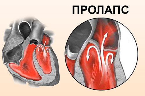
Among all heart defects, prolapse of the mitral valve is quite common. The disease is of three degrees of severity, and the most favorable prognosis is given by the prolapse of the mitral valve of the 1st degree. For proper treatment and prevention of disease, its symptoms should be properly identified.
