

What heart examinations do you have?
Author Ольга Кияница
2017-10-30
Heart tests help determine cardiovascular disease of varying degrees of severity. If previously used only by physical examination, today the most diverse methods of research and advanced technologies are used.
A timely examination of the heart reduces the risk of developing cardiovascular disease, which ranked first among the main causes of mortality.
Every patient entering the cardiology department or visiting a doctor in the clinic due to heart disease undergoes a standard research complex. If after that it is not possible to establish the root cause of the pathology, then narrow-minded methods of diagnostics are used.
Video Worrying Heart? Where to start the survey?
Primary examination of the patient's heart
The very first method of diagnostics of a patient with cardiovascular pathology is a physical examination at the first appointment of the doctor. Initially, an examination is conducted in order to detect visual changes (swelling, cyanosis, throat). After using the tapping, the doctor determines the limits of the heart, which in pathological cases change.
Auscultation of the heart is an important method of physical examination by which pathological rhythms and noises are determined, cardiac tone is expressed.
The folodoscope is used for auscultation. With the same tool combined with a tonometer, the blood pressure is measured. In the process of listening to the heart tones, their sound, and the order of interest are studied. In normal condition, two permanent tones, the first and the second, are heard. For this or that pathology, additional, as well as the third and fourth tones may be detected.
Video Basic physical examination methods of the heart
Instrumental diagnostic methods
Progressive technologies allow today to carry out the most complex and previously unavailable research. It is extremely important to prescribe proper treatment, since the exact diagnosis is the pledge of effective therapy. As a rule, start with standard methods - electrocardiography, ultrasound of the heart. If necessary, the examination is supplemented by electrophysiological examination, MRI, CT, angiocardiography.
Commonly used diagnostic tools:
- Electrocardiography.
- Echocardiography.
- Angiocardiography.
Part of these methods is a painless technique, while others, on the other hand, relate to invasive diagnostics. But all without exception are carried out with the help of special equipment, which, to a greater or lesser degree, affects the human body.
Electrocardiography
It is a valuable method of research in cardiology, by which most of the cardiovascular diseases are diagnosed. For the first time, the electrocardiogram was shot by Willem Einthoven, who developed a string galvanometer, which is practically the same ECG recorded in the twentieth century, as it is today. The same investigator developed a system of notation for the resulting teeth, which is still widely used.
Registering the electrical activity of the heart is done using an electrocardiogram, which is used for writing thermal paper today. If the device is completely electronic, then the received data can be stored in the computer.
Electrical indices are determined with the help of electrodes, which on the electrocardiogram show the potential difference in the form of teeth. For their production, standard leads (I, II and III) are used, which, according to charges, are superimposed on the right arm and the left leg (+) and the left hand (-). Also recorded are reinforced leads, denoted as aVR, aVL, aVF, which are derived from the extremities. In addition to these six leads, one-pole thoracic leads, from V1 to V9, are recorded, but most often V1-V6 is determined.
An electrocardiogram allows you to determine:
- scar changes;
- blood supply disturbance;
- dystrophic manifestations;
- signs of myocardial infarction;
- signs of rhythm disturbance.
The standard ECG does not always allow you to accurately detect pathological changes in the heart, so if necessary, other studies based on electrocardiography are used:
- Daily ECG monitoring - Defines mild heart rhythm disturbances that are difficult to fix with a standard ECG. The patient needs to wear a portable electrocardiogram from day to day, up to 5-7 days, which records the activity of the heart, after which the physician conducts an analysis of the data received.
- ECG mapping, or precardive mapping - when using a large number of electrodes, a long study is carried out, resulting in even the most difficult and difficult to diagnose the disease can be successfully identified. All the information received by the device is processed by the computer, therefore high accurate results are obtained.
- Load texts (bicycle ergometry, treadmill test) - are performed to determine heart lesions that are not detected in normal conditions. When performing physical activity, favorable conditions are often created for arrhythmias or other cardiac disorders. In particular, using bicycle ergometry is determined stress angina, ischemic heart disease, etc. When re-examination can be found tolerance to physical activity, the effectiveness of previously performed treatment, predictive value.
- Intraperitoneal electrocardiography - an active electrode is inserted into the esophagus, which is brought to the heart as close as possible. With the help of this method it is possible to evaluate the efficiency of the atrioventricular compound and the atrium. It is often used at the diagnostic stage for various types of rhythm disturbances, especially cardiac blockade.
- Vectorcardiography - when using the projection chart, a volumetric figure is formed that reflects the electrical activity of the heart. If there is a violation of the rhythm, the corresponding changes in the cardiac activity vector are recorded.
- Gastrocardiomonitoring is a method of simultaneous examination of the electrical activity of the heart and acidity in the stomach and esophagus. According to the method of conducting similar is not Holter monitoring, only during the day is recorded not only the electrocardiogram, but also pH-metry. The examination is often used in the diagnosis of gastrointestinal and cardiovascular diseases.
Electrocardiography is one of the safest and easiest methods for research. It is available at any level of medical care, so if you want and you can get a lot of reliable information about the state of the heart.
Video Heart research methods. ECG and PKG
Echocardiography
This method is more commonly known as ultrasound, or ultrasound of the heart. The principle of the study is based on the capture of signals that have been reflected from various heart structures. Depending on the acoustic density, the signal is perceived differently, but in the end the corresponding image is formed.
It is used to detect organic heart lesions, congenital and acquired vices, as well as to evaluate the functionality of the myocardium. It is considered a method of choice when examining people with complaints of weakness, dizziness, pain in the heart, loss of consciousness, frequent heartbeat.
On the basis of ultrasound, a number of modified heart tests have been developed:
- Doppler-echocardiography - on the ultrasound monitor, intracardiac blood flow is shown, which allows you to see heart defects, pathological shunts and chords, and to evaluate hemodynamics of the heart.
- One-dimensional echocardiography - allows you to see the heart in one plane. Pretty rough diagnostics, which is used to obtain the size of the organ, the thickness of its walls. Also, data are available on the operation of the valve apparatus, contractility of the heart.
- Two-dimensional echocardiography is a more informative study compared to the previous one. This is achieved by obtaining a solid image of the heart and its structures.
- Stress-ECHO is one of the options for load tests. Combines ultrasound and bicycle ergometry techniques. First, an ultrasound diagnosis is performed, and after the "riding" of the patient on a bicycle, an ultrasound is performed. As a result, it is determined by coronary heart disease, coronary vascular obstruction, and the effectiveness of therapy is evaluated.
No ultrasound is prescribed in cases of chronic smoking, bronchial asthma, in the presence of large mammary glands or excessive breast enlargement of the breast. Also, the study is not conducted with chest strains, infectious diseases of the skin in this area.
Echocardiography refers to harmless and painless methods of investigation. Performs quite quickly and, if necessary, repeats the required number of times.
Video echocardiography, ultrasound of the heart, research method
Angiocardiography
The method relates to X-ray examination, in which the X-ray contrast agent is used. With the help of the study it is possible to study the heart chambers, as well as the nearest veins and arteries.
A radiopaque substance is introduced into the cavity of the heart and coronary vessels, for which a special catheter is used. It is fed to the heart and vessels through the femoral or subclavian artery. As a result of the introduction of the contrast agent, the structural parts of the heart become visible, for the evaluation of which a number of images are made. Before performing the procedure, it is obligatory to take a sedative and antihistamines medicine.
The study is often performed prior to heart surgery in order to clarify the required physiological parameters of the myocardium. Also, the method is quite effective in the diagnosis of heart disease, disturbance of the structure of the nearest large vessels. After widespread echocardiography, angiocardiography has become less commonly used. But in some cases, this method is irreplaceable, since it allows more accurately to determine the anatomical structures of the heart.
Angiocardiography refers to invasive diagnostic methods, therefore relatively infrequently used. Nevertheless, if necessary, it helps to obtain more accurate data than with echocardiography.
Video Angiography and stenting of the vessels of the heart
Laboratory analyzes
They are often prescribed if there is a high risk of co-morbidity. Also, with certain heart defects in the blood, certain substances are detected, fixed with the help of special laboratory tests.
Common laboratory tests for heart disease:
- Urine tests - the kidneys react sensitively to the condition of the cardiovascular system. In edemas, after the onset of paroxysmal tachycardia, septic endocarditis, the quality and quantity of urine varies. Basically, the violations are expressed in the decrease in specific gravity of the urine, the determination of hyaline cylinders, red blood cells, protein in the urine.
- A blood test is quite informative in the diagnosis of cardiovascular disease. In particular, often with heart defects increases the number of red blood cells. A similar change is associated with oxygen starvation with cardiac insufficiency. Inflammatory processes often affect endocardium, myocardium and other structures of the heart. At the same time leukocytosis develops and ESR increases.
- Sputum examination - is conducted only in cases where there is suspicion of acute failure of the left ventricle. With this pathology, stagnation in the lungs is observed, resulting in bloody foamy sputum. In some cases, it may be inflammatory, which is often noted with pronounced congestion of the lungs. Microscopic analysis of sputum allows to define "cells of cardiac cages", the presence of which is characteristic for myocardial infarction and heart failure.
During each study, it is required to carefully follow the requirements of the doctor conducting the diagnosis. This will help you get a faster and better result. Also, subsequently, it will prevent the development of complications, therefore the joint cooperation between the doctor and the patient helps to achieve the desired result.
Similar articles
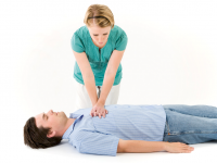
In critical situations, when a person does not have a heart rhythm or respiration, cardiopulmonary resuscitation is performed first. With the help of standard techniques, you can help the patient avoid death, or at least support his life activity until the arrival of the medical staff.

The heart belongs to vital organs, therefore without it it would not have been possible for a single person to exist. The structure, as well as the appearance of the heart, is rather diverse, but quite capable of logical explanations. A proper understanding of how the human heart looks like a healthy estimate of the body’s overall capacity.

ECG (or electrocardiogram) is one of the most common and accessible research methods and demonstrates the details of the functioning of the heart in the form of a graphic image that reflects the electrical activity of the organ. This method is often used for the initial diagnosis of cardiac pathologies, is carried out as part of a comprehensive examination of the patient before presumed treatment or during preventive examinations.