

What does sinus tachycardia mean in children of different ages?
Author Ольга Кияница
2018-07-02
Sinus tachycardia (CT) is a change in the rhythm of the heart toward its increase, in which the sinus node remains the main rhythm driver. In children, this condition is very common, which is often associated with both their increased activity and rather rapid growth.
In pathological cases, tachycardia is observed in a calm state, and in addition to rapid heart rate, other pathological signs are determined.
Diagnosed tachycardia is simple, for which it is enough to correctly calculate the heart rate on the radial or carotid artery. To determine the type of tachycardia, electrocardiography is required. If the rhythm disturbance affects the child's well-being, then the treatment is not carried out. Otherwise, additional diagnostics are performed and appropriate therapy is then given.
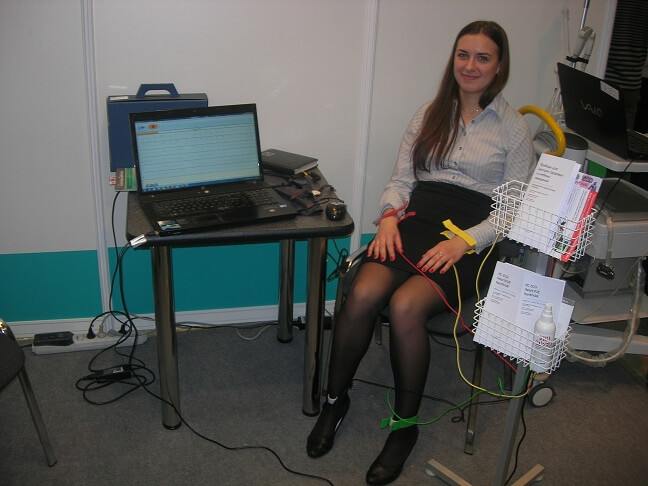
Video: Sinus tachycardia in a child
Description of sinus tachycardia
Sinus tachycardia is one of the types of arrhythmias, in which the heart rate is more than typical for a particular child's age. This is due to the change in the operation of the sinus node, which sends electrical impulses at a higher rate than usual.
In the normal state, the heart rhythm is set by the first-order driver - the sinus node, therefore, when only one indicator (HR) changes and no clinical manifestations, sinus tachycardia is defined as a variant of the norm.
If the heart rate becomes too fast, the circulatory system is disrupted in the body. In such cases, CT can cause the following symptoms:
- weakness;
- fatigue;
- dizziness;
- a feeling of palpitations.
In small children, CT can be manifested by fussiness, difficulty feeding, capriciousness, excessive excitation, changes in the coloration of the skin, etc.
Sinus tachycardia is often temporary, when the body experiences stress from physical exertion, intense emotions, fever, or dehydration. Sometimes the child is affected by several reasons. As soon as the external factor of exposure is eliminated, the heart rate usually returns to normal.

Sinus tachycardia and age of the child
In an adult human heart rate is 60-90 beats per minute. In children, the indicators are somewhat different, with the smaller the age of the child, the more often the heart is contracted.
Table of normal values of heart rate in children of various ages
| Age | Normal heart rate (Average value) (bpm) |
| up to 1 day | 93-154 (123) |
| 1-2 days | 91-159 (123) |
| 3-6 days | 91-166 (129) |
| 1-3 weeks | 107-182 (148) |
| 1-2 months | 121-179 (149) |
| 3-5 months | 106-186 (141) |
| 6-11 months | 109-169 (134) |
| 1-2 years | 89-151 (119) |
| 3-4 years | 73-137 (108) |
| 5-7 years | 65-133 (100) |
| 8-11 years old | 62-130 (91) |
| 12-15 years old | 80-119 (85) |
| over 16 years old | 60-100 |
As can be seen from the table, only from the age of 16 years in children the norm of the heart rate becomes the same as in adults.
Looking at the table, you can immediately determine whether there is a tachycardia in the child. For this, the pulse is calculated for one minute, then according to age, the proper range is determined. If in fact the heart rate is greater than the upper value indicated in the table, then the child has tachycardia.
With sinus tachycardia, the rhythm of the heart remains correct, although it is rapid, so additional diagnostic methods are used to determine the type of arrhythmia.
Causes of sinus tachycardia in children
There are various factors of influence that can cause in childhood tachycardia. Most often, the ST appears again against the background of the following states:
- Dehydration / hypovolaemia (most often)
- Fever
- Hypoxia
- Anemia
- Shoka
- Pulmonary edema
- Hyperthyroidism
- Hypocalcemia
Separately, it should be pointed out that sinus tachycardia can cause improper intake of medications. As children are very fond of trying everything new, it is their familiarity with medicines in the absence of adult supervision that can lead not only to ST, but also more severe consequences.
Drugs and substances that cause tachycardia
- amphetamines;
- phenothiazines;
- caffeine;
- chloride hydrate;
- antihistamines;
- phenothiazines;
- antidepressants;
- tobacco;
- theophylline.
Diagnostics
During the examination of the child, the doctor may ask the following questions:
- Does the child have a history of the disease, in particular sinus tachycardia, or is there some other kind of heart disease?
- What was the beginning and duration of the current illness in the child?
- Does the child have other symptoms, such as:
- chest pain;
- inconsistent breathing;
- discoloration of the skin;
- loss of consciousness;
- neurological symptoms (for example, a change in the level of consciousness);
- Does the child take any medications?
- Does the child have anything to do with an allergy?
- Is there a family history of heart disease?
- Is there a tachycardia at a particular time or under certain circumstances (for example, with activity, after eating, under stress)?
- Did the child complain about the feeling of fever / fever?
- Did the child have vomiting or diarrhea?
- How often does the child drink and pee?
In addition to the complete medical history and physical examination of the child, various types of procedures are performed that can be used to diagnose CT. In particular, the following methods are used:
- Electrocardiography (ECG) is a measurement of the electrical activity of the heart. When the electrodes are located in certain places on the body (chest, arms and legs), a graphic image is created in the form of teeth of different heights and shapes. ECG may indicate the presence of arrhythmia or other types of heart disease.

There are various variants of the ECG test, including:
- Resting ECG (ECG at rest) - to carry out this study, the child is stripped to the waist, and after the doctor attaches electrodes in the form of small pads to the chest, shoulders and legs. On the other hand, the electrodes are connected to the ECG apparatus. The device starts and registers the electrical activity of the heart for a minute or so.During this, the child lies quietly.
- Stress test . The child joins the measuring equipment, as described above. However, instead of lying, the child does the exercise, moving on the treadmill or "driving" on a stationary bike. During the movement, an ECG is recorded.This test is carried out to assess the performance of the heart during exercise, which is partly consistent with the level of emotional stress.
- ECG of high resolution . This procedure is performed in the same way as the ECG at rest, except that the electrical activity of the heart is recorded for a longer period of time, usually 15 to 20 minutes. The method is used for the suspicion of arrhythmia, which is not fixed by the standard ECG, since rhythm disturbances can be short-lived in nature and do not occur during recording in the usual way.
- Holter monitoring is an ECG record that is performed for 24 or more hours. For the study, three electrodes are attached to the baby's chest and connect them to a small portable ECG recorder using wires. The child conducts his usual daily activities (with the exception of such actions as showering, swimming or any work that causes excessive sweating, because of which the electrodes may fall off). There are two types of Holter monitoring:
- Continuous recording - ECG is recorded continuously throughout the testing period.
- Event monitor - The ECG is recorded only when the patient begins a palpitations.
- Electrophysiological research (EFI) is an invasive method of diagnosis, in which a small thin tube (catheter) is inserted through the vessel into the groin or neck and advances to the heart. This gives the doctor an opportunity to find the place of arrhythmia directly from the heart cavity, additionally determining how to best treat.
Differential diagnosis of sinus tachycardia with other types of arrhythmia
| Sinus tachycardia | Supraventricular tachycardia (reentry) | Atrial fibrillation | |
| Clinic | May be absent, or fever / sepsis / shock / anemia |
50% - Wolff-Parkinson-White syndrome 25% - atrioventricular, nodular, reentral tachycardia, Ebstein abnormality |
90% have an enlarged atrium / myocarditis / intoxication with digoxin |
| Heart rate | Usually <200 / min | Infants: <300 / min, older children: <240 / min. | Atrial frequency 250-400 / min 2: 1, 3: 1 or 4: 1 with AV block |
| Zubets P | Normal | It can be hidden in the QRS complex, in 60% there is a retrograde P-wave | Regular flutter waves (F-waves) |
| QRS | Normal | Normal or aberrant | Normal |
Thus, sinus tachycardia is most clinically beneficial, since other forms of arrhythmia are often accompanied by more severe and severe clinical symptoms.
Treatment
The specific treatment of sinus tachycardia will be determined by the child's attending physician on the basis of:
- Age, general health and medical history
- Degrees of clinical manifestations
- Tolerance of the child's organism to specific medicines, procedures or therapies
Sinus tachycardia may be present, but it is almost impossible to manifest and not cause any health problems. In this case, the doctor can recommend only to adjust the lifestyle, if required. However, when a rhythm disorder causes symptoms, there are several different treatment options.
Typically, the doctor chooses the treatment strategy according to the type of arrhythmia, the severity of the symptoms, and the presence of other pathological conditions (for example, diabetes, renal failure, heart failure, thyroid dysfunction) that may affect the course of treatment.

Most often sinus tachycardia can be treated by changing the lifestyle and medicines. In other cases, the following methods of therapy are used:
- Cardioversion . This procedure is based on the effect of a weak electrical voltage delivered to the heart through the thorax, which allows stopping certain very fast arrhythmias such as atrial fibrillation, supraventricular tachycardia, or sinus tachycardia. Initially, the child is given a medicine to help him relax, and then connect the monitor to the ECG, which is in parallel with the cardioversion device. A weak electric current is applied to a certain point of the heart during the ECG cycle.
- Ablation is an invasive procedure performed in the electrophysiotherapy department. It is carried out by means of a small thin tube (catheter), which is injected into the heart through the vessel into the groin or hands. The procedure is similar to the electrophysiological study described above. Once the causative site of the arrhythmia is determined by EFI, the catheter is moved to it. The cause of rhythm disturbance can be destroyed by using the appropriate technique:
- Radiofrequency ablation (the site of the lesion is exposed to very high frequency radio waves, which heat the tissues before they are destroyed)
- Cryoablation (part of the myocardium is exposed to ultra-cold matter, freezing tissue and then destroying them).
- Implantation of a cardioverter-defibrillator (ICD) . A device similar to a pacemaker is implanted near the heart.When the heartbeat becomes too fast, the device generates a weak electric current that acts on the heart and, consequently, slows its rhythm.
- A pacemaker is a small device that is implanted under the skin and sends electrical signals to start or correct a slow heartbeat. A permanent pacemaker can be used to heart beat if the natural heart pacemaker (sinoatrial node) does not function properly. With this indication can be not only abnormal heart rhythm, but also blocking the conduct of electrical impulses. Pacemakers are usually used in the treatment of delayed rhythm by the type of sinus bradycardia, weakness syndrome of the sinus node or cardiac blockade.
In infants and small children, pacemakers are usually placed in the abdominal cavity. The wires that connect the pacemaker to the heart are placed on the outer surface of the heart. This precaution is necessary because the fat layer in the abdominal cavity protects the pacemaker wires and the device itself from impacts that can occur during daily activities in childhood, for example, when lifting or falling.
School-age children and adolescents, a pacemaker can be installed in the shoulder area under the collarbone.Pacemaker wires are often placed inside the superior vena cava, a large vein that connects to the right atrium, and then travels to the heart.
Surgical intervention is usually performed only when all other modern therapies have not produced the expected result.Surgical ablation is the main surgical procedure that requires general anesthesia. The thorax is cut and the operation is performed on the open heart. The place of an arrhythmic focus is established, then it is destroyed or removed, which allows to eliminate the arrhythmia.
Video: What is heart tachycardia and how to treat it?
Forecast
With sinus tachycardia, a prognostic opinion is most often favorable. It is enough to follow medical recommendations to improve the child's condition.
If a marked clinical clinic is observed or the child has a concomitant cardiovascular disease, then the prognosis worsens and much depends on the effectiveness of the chosen therapy strategy.
Similar articles

The process of pacemaker installation is not as complicated as it may seem. Today, this manipulation is equated to an operation to remove appendicitis. More questions arise in patients how to live, after the installation of ACS, but the process of surgical intervention itself is important.
Modern technologies allow the most thorough study of various organs and tissues, including the cardiovascular system. Without timely and correct diagnosis sometimes it is very difficult to put the exact diagnosis. At the same time, it is extremely important to do everything in time to ensure effective treatment after the study.
Prevention of the development of cardiovascular diseases is done by preventive cardiology. To date, various schemes have been developed to prevent such formidable diseases as myocardial infarction, heart rhythm disturbance, stroke, heart failure. Timely measures taken will prolong life.